MissionVet Specialty & Emergency
MissionVet Specialty & Emergency treats a wide variety of orthopedic and soft tissue surgical diseases. MissionVet is staffed by board-certified veterinary surgeons that specialize in state-of-the-art surgical treatment for your pet.
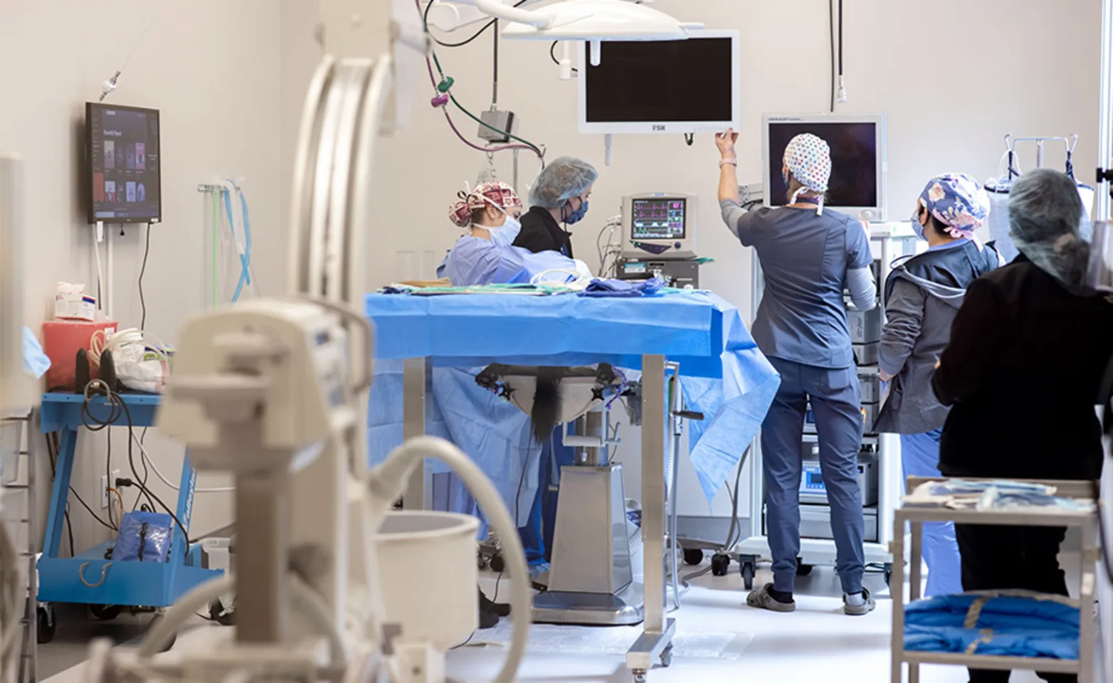
Orthopedic surgery encompasses a wide range of joint and bone surgery. One of the more exciting and innovative aspects of orthopedic surgery is the ability to perform arthroscopy. Just as with people, arthroscopy allows us to visualize your pet’s joints with small cameras. Because of the smaller incision, your pet can go home sooner and be more comfortable. In addition, the high-definition camera gives a better view of the joint than the naked eye so that no abnormalities are missed. Treatment of cruciate and meniscal tears as well as elbow dysplasia is commonly performed with arthroscopy allowing for only 2-4mm (1/4 inch) skin incisions.
In addition to joint disease, Orthopedic surgery also focuses on the treatment of fractured bones. Using specialized bone plates or external fixators, we repair all forelimb, hindlimb, and pelvic fractures. In addition, we work closely with our board-certified Neurosurgeon on spinal fracture repairs.
Shoulder Disorders
The most common condition that is treated with the shoulder of dogs is Osteochondritis Dessicans (OCD). This is often found in young, large breed dogs and is caused by an abnormality of the growth of the cartilage within the shoulder joint. This abnormal growth causes the cartilage to become thickened and eventually loosen from the underlying bone creating a flap of cartilage. This condition is treated by removing the fragment and debriding the underlying bone to allow fibrocartilage formation. This procedure is most often performed arthroscopically which decreases surgical time, post-operative pain, and recovery time. Most patients can be discharged the same day as surgery. There is a very good prognosis for patients treated for shoulder OCD. Biceps tenosynovitis is also a painful condition of the shoulder that can be diagnosed and treated arthroscopically. It may result from trauma to the joint or from a soft tissue injury after strenuous activity. It is typically painful on palpation over the biceps tendon and can often be diagnosed on physical examination.
Fracture Repair
Fractures (broken bones) are often suffered by dogs and cats after some degree of trauma (falls, hit-by-car, etc.). The most common bones affected are the radius, tibia, humerus, femur, and pelvis. Some fractures in young animals can be treated with a cast or splint for several weeks. More complex fractures in adults often require surgical stabilization. This can be accomplished by numerous methods including plate/screw fixation, pins, wire, interlocking nail, and linear or circular external fixation. The method of repair will be determined by the location and severity of the fracture as well as patient and owner factors. We also have the capability of intra-operative fluoroscopy which allows us to evaluate the repair in real-time. This decreases surgical time which in turn decreases the infection rate and decreases the anesthetic recovery time for the patients. Fluoroscopy also allows for minimally invasive plate osteosynthesis (MIPO) which can speed the healing time of the fracture. Healing time for most fractures ranges from 4-6 weeks in young animals to 8-10 weeks in adult animals.
PennHIP
PennHIP is a radiographic screening method used by PennHIP-certified veterinarians to assess, measure and interpret hip joint status. Performed on dogs as young as 16 weeks of age, PennHIP’s primary objective is to reduce the frequency and severity of hip dysplasia in all breeds of dogs. Stored in and compared to an extensive collection of PennHIP data containing over 100,000 patients, PennHIP x-rays allow pet owners the ability to obtain an estimate of their dog’s risk for developing hip dysplasia and, if necessary, make lifestyle adjustments for their dog to enhance the quality of their pet’s life. PennHIP x-rays also allow breeders to confidently identify the members of their breeding stock with the best breeding potential. PennHIP was established in 1993 after 10 years of research by Dr. Gail Smith in evaluating a technique that quantified hip laxity. PennHIP has published more than 40 manuscripts over the past 20 years.
Arthrodesis
Arthrodesis is another term for the fusion of a joint. This is needed when there is significant damage to a joint, either from trauma or from severe arthritis. It prevents the joint from moving and therefore eliminates the pain associated with the painful condition in that joint. Depending on the condition the entire joint may need to be fused (pan-’joint’ arthrodesis) or only part of the joint may need to be fused (partial ‘joint’ arthrodesis).The most common joints that are fused are the carpus (wrist) and tarsus (ankle). The shoulder, elbow, and knee can also be fused but the need is much less common.
Cranial Cruciate Ligament Rupture (CrCLR)
This is the most common cause for a hind limb lameness in dogs. There are many theories as to why this ligament tears in dogs, but a single definitive cause has not been identified. We do know that the ligament slowly degenerates and tears with time and often can completely tear with minimal to no trauma. Approximately 40-60% of dogs that have a cruciate tear in 1 knee will have the other one tear at some point in their life. The main sign that is noted is limping in the affected leg. This can be a severe as not placing any weight on the limb or as mild as shifting weight off of the leg when standing. Cruciate tears can be diagnosed with a combination of physical exam findings as well as radiographs (x-rays) of the knee. The diagnosis is confirmed with an arthroscopic examination of the cruciate ligament at the time of surgery. Using arthroscopy as opposed to arthrotomy increases visualization of the joint, decreases post-operative pain, and increases patient use of the operated limb which speeds post-operative healing. There are several surgical treatment options for dogs with cruciate tears. The one that has shown to have the best short and long term outcome is the Tibial Plateau Leveling Osteotomy (TPLO). This is a procedure that decreases the tibial slope to a point that eliminates the abnormal motion in the knee caused by the cruciate tear.
For dogs that have severe arthritis in the knee that is no longer responsive to medical management, we offer total knee replacements. This is a procedure that replaces both the femoral and the tibial components of the knee joint. It is very similar to the procedure that is performed in people for knee replacements.
Medial Patellar Luxation (MPL)
Patellar luxation is a common finding in young small breed dogs but can also be found in large breed dogs. Even though your pet may have patellar luxation it may or may not require surgery. We recommend correction of patellar luxation in patients that are experiencing pain and lameness secondary to the luxation. You may notice that your dog skips on the affected leg or holds it up when running or walking. They may also stretch the leg backwards in order to replace the patella back in its normal location. There are 4 procedures that are most often required to correct the luxation. The normal groove that contains the patella often has to be deepened (trochleoplasty) to maintain the patella in the proper location. The attachment of the patellar tendon (tibial tuberosity) must be realigned with the remainder of the limb by cutting the bone and moving the tuberosity to a more normal location. When the patellar luxation occurs toward the medial (inward) of the knee this tissue must be released (retinacular release) to allow the patella to return to its normal location and the tissue on the lateral (outward) side of the knee must be tightened (fascial imbrication) to keep the patella in place.
Hip Dysplasia
Hip dysplasia is a common problem diagnosed in mainly large breed dogs. It is a developmental disorder that starts with increased laxity in the hip joints which leads to abnormal articulation and wear of the joint surfaces. This leads to the formation of arthritis as the dog ages which can cause it to be painful and difficult for the dog to rise, run, jump, and play like they did before. There are surgical options for hip dysplasia depending on the age of the dog. Juvenile Pubic Symphysiodesis (JPS) is a procedure that is designed for dogs between 16 weeks and 20 weeks of age. It causes a portion of the pelvis to be fused in a growing dog which causes the pelvis to grow in a way that actually increases the “tightness” of the joint. This decreases the wear of the joint and decreases the risk of arthritis in the future. Double Pelvic Osteotomy (DPO) is a procedure that is performed in young dogs (<1yr) that have laxity but no evidence of arthritis and relatively normal hip conformation. Two cuts are made in the pelvic bone that allow the acetabulum (cup) to be rotated over the femoral head, increasing the coverage and decreasing the progression of arthritis in the future. The best option for dogs with hip arthritis is a total hip replacement (THR). This procedure replaces the acetabulum (cup) as well as the femoral head (ball) to eliminate the pain associated with the cartilage damage and arthritis that has developed. A total hip replacement is as close to a normal hip as possible and can allow dogs to return to normal function after the post-operative recovery period. The second option for dogs with severe hip arthritis is a Femoral Head and Neck Ostectomy (FHO). This is a procedure where the femoral head and neck are removed from the socket joint. Scar tissue forms a “false joint” that eliminates the bone on bone contact associated with severe arthritis. This procedure is better tolerated in cats and small dogs (<20 pounds). Hip luxations can also be treated with an FHO or total hip replacement. More commonly we treat hip luxations with a Toggle Pin repair technique that allows your dog to maintain their own hip joint.
Elbow Dysplasia
Elbow dysplasia is a condition commonly diagnosed in large breed dogs (Labrador retriever, Golden retriever, Rottweiler, German Shepherds, Newfoundland, Bernese Mountain dogs) and is composed of several different components (Medial Coronoid Disease (MCD), Osteochondritis Dessicans (OCD), and Ununited Anconeal Process (UAP). Dogs can be affected by one or multiple components of this condition. In most cases it can be treated arthroscopically which minimizes surgical trauma associated with a large incision into the joint and decreases post-operative pain and recovery time. The elbow is a very tight and congruent joint and any abnormality present can lead to the progression of arthritis. Some dogs diagnosed with this condition will require treatment of arthritis as the dog ages. This may be as simple as giving fish oil and joint supplements or as complex as requiring joint injections and rehab therapy.
Cranial Cruciate Ligament Ruptures in Dogs
Learn more about the causes, diagnosis, and treatment of cranial cruciate ligament tears in dogs, and gain a better understanding of the TPLO surgery used to stabilize the knee when a tear occurs.
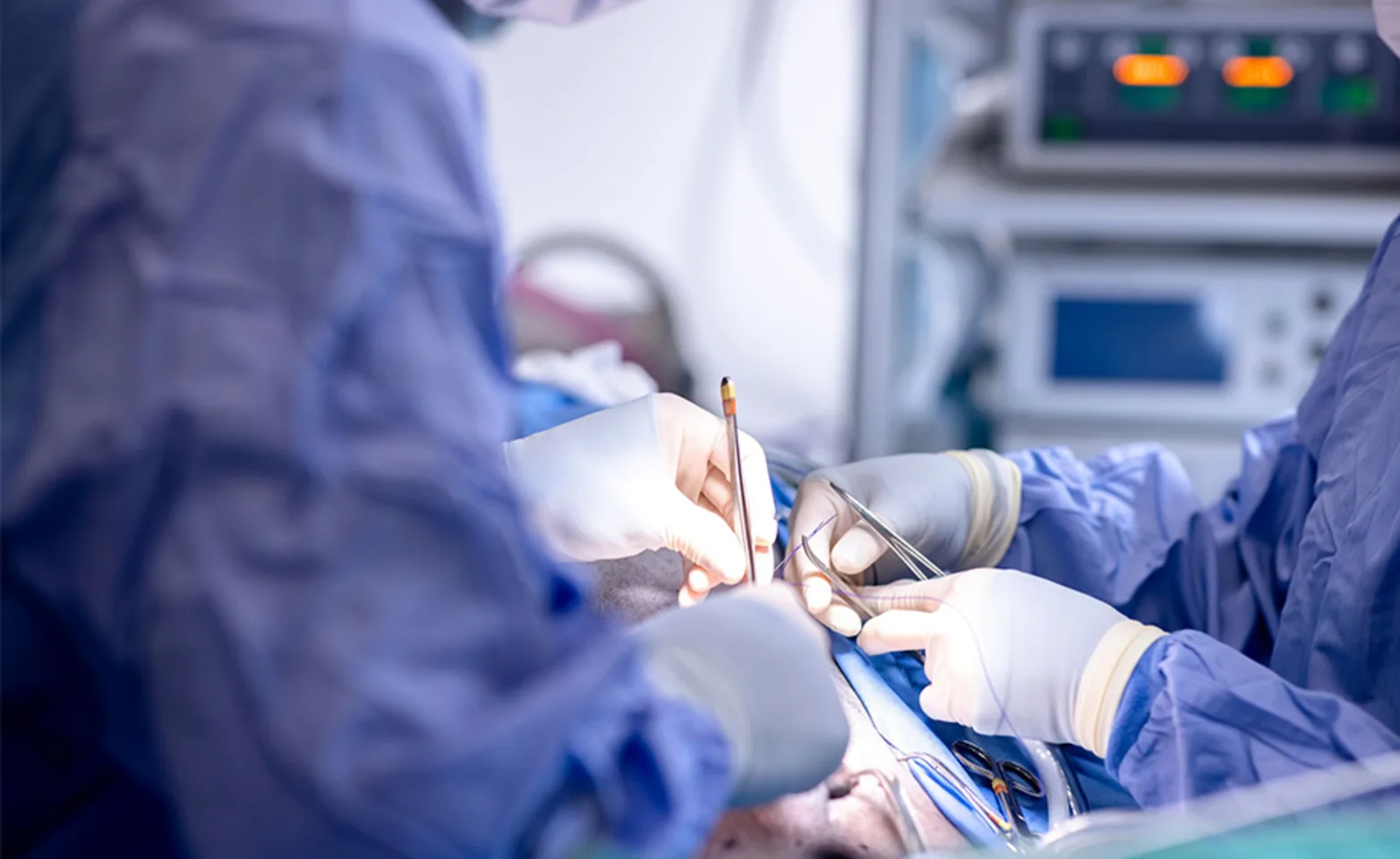
Soft tissue surgery includes a variety of surgical treatments from foreign body removal to hernia repair to organ biopsies. We work with traumatic wounds (from dog fights to animals hit by cars) as well as oncologic surgery (including mass removals and amputations). Your pet’s comfort and welfare are always paramount and we treat each pet as if they were our own. We even have the capability to perform Minimally Invasive Surgery on select cases.
Wound Care and Reconstruction
We offer state of the art wound management including standard open wound management and vacuum assisted closure (VAC). The VAC wound management system can be used for large open wounds or for chronic wounds associated with areas that are difficult to heal. In some instances skin grafts or skin flaps are needed to close wounds. The VAC bandage is often used after these procedures as well to increase survival of the skin graft or flap by decreasing motion and increasing blood supply to the skin. Based on each particular wound your pet’s surgeon will discuss the different options and recommend the best management for your pet.
Abdominal Surgery
Abdominal surgery consists of procedures on numerous different organs within the abdomen. These include the liver, gall bladder, stomach, small intestines, colon, pancreas, adrenal glands, bladder, urethra, kidneys, and ureters. Some of these procedures may be able to be performed minimally invasively with laparoscopy to decrease post-operative pain and the recovery time associated with the procedure.
Hernia
Diaphragmatic hernia
Body wall hernia
Hiatal hernia
Peritoneal-pericardial hernia
Perineal hernia
Liver
Liver lobectomy for abscess or tumor removal
Liver biopsy (open or laparoscopic)
Portosystemic shunts
Gall bladder
Cholecystectomy
Cholecystoenterostomy
Stomach
Gastropexy (open or laparoscopic)
Gastrotomy (foreign body)
Gastric Biopsy
Gastric Dilation and Volvulus (GDV)
Small intestines
Biopsy
Enterotomy or Resection and Anastomosis for foreign body removal,
Tumor removal
Colon
Tumor resection
Megacolon
Pancreas
Biopsy
Tumor Resection
Abscess Drainage
Adrenal glands
Adrenal mass removal, Adrenalectomy
Bladder
Cystotomy (bladder stones)
Biopsy
Prostate
Prostatic biopsy
Prostatic abscess
Prostatic cysts
Urethra
Urethrotomy (urethral stones)
Perineal urethrostomy (Feline Lower Urinary Tract Disease)
Kidneys
Nephrectomy (tumor, trauma)
Nephrotomy (Kidney stones)
Ureter
Ureterotomy (ureteral stones)
Neoureterocystostomy (ectopic ureter)
Ovaries and Uterus
Ovariohysterectomy (pyometra)
Ovariectomy (open or laparoscopic)
Perianal-anal sacculectomy
Perianal mass removal
Thoracic Surgery
Thoracic surgery consists of procedures on numerous different organs within the thoracic cavity. These include the lungs, heart, great vessels, and general thoracic cavity. Some of these procedures may be able to be performed minimally invasively with laparoscopy to decrease post-operative pain and the recovery time associated with the procedure.
Lungs
Lung lobectomy- tumor
Foreign body
Lung biopsy
Heart
Heart based mass resection
Pericardectomy
Great vessels
Patent Ductus Arteriosus (PDA)
Persistent Right Aortic Arch (PRAA)
Thoracic cavity
Chylothorax
Pyothorax
Mediastinal tumor resection
Upper Airway Surgery
Upper airway surgery consists of surgical procedures performed on the larynx, the trachea, nose, and soft palate. The most common upper airway conditions that are treated with surgery are brachycephalic syndrome (English bulldogs, Pugs, French Bulldogs, etc.), laryngeal paralysis (Labrador retrievers), tracheal collapse (small and toy breed dogs).
Brachycephalic syndrome
Elongated soft palate resection
Opening stenotic nares
Removal of everted saccules
Laryngeal paralysis
Arytenoid lateralization (Laryngeal tie-back)
Vocal fold resection
Tracheal collapse
Prosthetic tracheal ring placement
Tracheal stenting
Surgical Oncology
Surgery is often only one component of treating cancer, which is why our surgeons work closely with our Oncology and Internal Medicine departments on patients diagnosed with cancer. Once we evaluate your pet we will make our treatment recommendations to allow the best care for your pet. Conditions treated can range from benign skin tumors, amputation of a digit or limb, to large malignant thoracic or abdominal tumors.
Head and Neck Procedures
Head and neck surgery consists of procedures performed on the ears, throat, and mouth of dogs and cats. Our dental surgery department performs surgery related to the mouth. Dogs and cats may also suffer from tumors in the neck (thyroid tumors) which may require surgery. The most common procedure performed on the ear is a total ear canal ablation (TECA) which is used to treat chronic ear infections. It eliminates any infected tissue and allows your pet to live a more comfortable life without the pain associated with chronic ear infections.
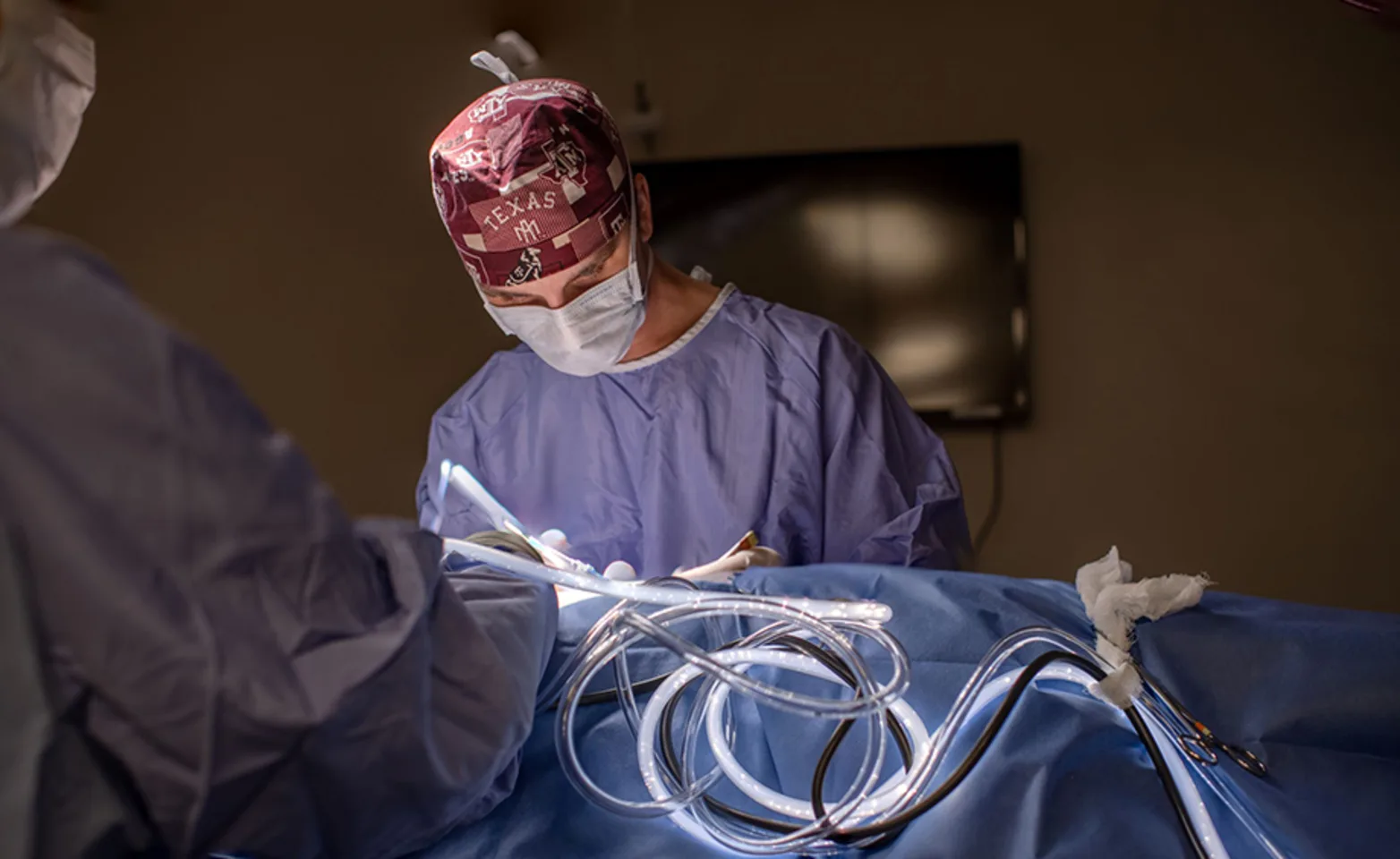
Minimally invasive surgery (MIS) is an exciting facet of surgery that allows for less pain and faster return to comfort for your pet. For MIS, we create small incisions through the skin to allow for access to the surgery site through skin ‘portals’. Through these portals, we can insert a small camera into different body cavities including the abdomen, chest and even joints. Though small incisions give a small working area, we are able to work with a magnified high-definition image that can often make the surgery easier and allow us to find problems that we might miss with a regular open surgery. Additional portals can easily be created to allow the surgeon to utilize specialized instruments to perform different procedures. The smaller incisions often allow for a faster recovery with less post-operative pain and a smaller incision for your pet.
Laparoscopy
Laparoscopy involves surgery with a small camera and scope placed in the abdomen. Laparoscopic surgeries include ovariectomy and liver biopsies.
Laparoscopy-Assisted Surgery
Laparoscopy-assisted surgery allows for small incisions into the abdominal wall (2-3 inches) to provide for easier access to abdominal organs without performing a full open approach. Laparoscopy-assisted surgeries include gastropexies (attachment of the stomach to the body wall for prevention of gastric dilatation and volvulus (GDV or ‘twisted stomach’) and ovariohysterectomies (spays).
Thoracoscopy
Thoracoscopy is like laparoscopy but allows for visualization and manipulation of organs in the chest. Thoracoscopic surgeries include biopsies of the lung and removal of the pericardium (sac surrounding the heart).
Arthroscopy
Arthroscopy utilizes a camera in joints with small portals for the placement of instruments. Arthroscopy can be used for the treatment of ACL and meniscal tears as well as elbow dysplasia.
Minimally Invasive FAQs
Does minimally invasive surgery cost more?
The simple answer is “yes, it does”. The specialized equipment cost, equipment maintenance, and disposable materials do add a significant cost to each procedure. However, we strongly feel that it is so important to provide the highest quality of care for our patients that we do not pass this cost along to you.
Why minimally invasive surgery?
As with the human surgical field, there is a growing trend in veterinary medicine to perform surgery in a minimally invasive manner. The popularity of minimally invasive surgery, or MIS, is due to the much smaller incisions that are needed; smaller incisions translate to less discomfort for your pet with a faster return to activity. Another big advantage is the increased visualization that can be achieved with MIS. Though the cameras are small, the images are projected on a large HD screen. Because the cameras are fitted with high-intensity xenon or halogen fiberoptic lights, visualization and magnification of the surgical site can be superior to conventional open surgery. Lastly, some procedures can be performed more quickly because of the decrease in time needed to close incisions.
Does it matter how big my pet is to have MIS?
Minimally invasive surgery can be performed in almost any size animal. Small puppies and kittens (less than 4 pounds) can be more difficult as there may not be enough room for the camera.
How long does it take to recover from surgery?
The recovery time after surgery depends on what procedure was performed. Usually your pet will be able to go home the next day; sometimes they can even go home the same day as surgery! We usually recommend 10 days of rest after surgery – though your pet often feels back to normal within the first day or two because the incision is so small. They will still have an incision that needs to heal and this generally takes 7-10 days.
Are there any risks or disadvantages to MIS?
The biggest risk of MIS is accidental puncture of the spleen during introduction of instruments into the abdomen. This can result in bleeding which is usually self-limiting; conversion to an open procedure is rare. We monitor patients very closely with all surgeries we perform, but with MIS, we pay particular attention to respiration as well fill the abdomen or chest with air while performing surgery. Because of this, we breathe for your pet so complications are rare.
The biggest disadvantage to MIS is that it can cost more to perform than conventional open surgery. The extra cost is due to the extra specialized equipment and training that is needed to perform the surgery. The cost is offset by decreased invasiveness and increased comfort to your pet, just as with human surgery.
Why do you recommend just ovary removal over ovary and uterus removal for sterilization with MIS?
Traditional spays in the United States are performed by removing both the ovaries and the uterus (also called ovariohysterectomy or OVH) of a female dog resulting in sterilization. Ovariectomy (or OVE) is removal of just the ovaries and is commonly done in European countries to sterilize female dogs and cats. While we commonly remove the uterus with conventional ‘open’ spays, it is much easier and faster and allows for a much smaller incision to remove only the ovaries with minimally invasive surgery. Numerous studies have shown that there is no increased risk of problems (such as pyometra or endometritis) if the uterus is left behind as ovary removal takes away all hormones that cause these problems.
FAQs
What do I need to bring to my pet’s appointment?
We recommend that you bring all medications (or a list of those medications) that your pet has been taking. In general, medications do NOT need to be stopped before your appointment. Also, please bring any records (blood work results, x-rays, etc.) that you have from your veterinarian – even if they already sent them over. It is always best to have copies with you in case the information does not make it here prior to your appointment.
If my pet stays in the hospital, when can I visit?
We know how hard it can be to be away from your pet. If your pet has to stay in our hospital, you can certainly visit. Our visitation hours are between 9 am and 5 pm, Monday through Friday. Please call ahead and we will arrange the best time to visit with your pet. We do not recommend visits the same day as surgery as we prefer for your pet to rest after their procedure and visits often get them excited.
When will my pet have surgery?
Your pet’s surgery will often occur on a separate day from your appointment – either the following day or a day that can be scheduled at your convenience in the future. Just as with human surgery, we use the initial appointment to perform a complete exam and any tests we may need prior to surgery. For some cases, however, we can perform the surgery the same day as the appointment so we recommend fasting your pet the night before (no food after 10 pm; water is okay).
What is my pet's recovery time?
Most pets stay one or two nights in the hospital before being discharged to go home. For soft tissue surgeries, we usually recommend 10-14 days of rest and confinement after surgery. For orthopedic surgeries, the recovery times depends on the surgery performed and can range from 2 to 10 weeks. The surgeon will make sure to go over the expected recovery time at the initial appointment.
What will I have to do after my pet comes home from surgery?
After surgery, you will need to monitor your pet’s incision at home. This entails just looking at the incision daily (if there is no bandage). We will sometimes send patients home with bandages that you can leave on until they fall off. If you cannot see the incision, you just need to monitor the bandage for strike-through. If you notice any drainage or discharge, please call us or your primary care veterinarian. We often send home E-collars to prevent your pet from licking at their incisions for the first 10-14 days (until the skin has healed). Although E-collars can be frustrating for both pet and owner, licking at the incision is very common for pets and is one of the main causes of post-operative infections – so it is so important to be strict with the E-collar. After orthopedic surgery, we usually recommend confinement to a small crate as this is as close as we can get to ‘bed rest’ for our pets. The crate should only be big enough for your pet to turn around and get comfortable but not stand up or jump around. For cats, their litter box should be placed in their cage and litter replaced with newspaper; for dogs, they should only go outside for short, controlled walks on a leash so they don’t run or play. While confining is difficult, it is critical to a good outcome for your pet and is one of the most important things you can do to help your pet recover faster and more comfortably.
Where do I go for recheck examinations?
We usually recommend a recheck of the surgical incision at 10-14 days. This can be done at your primary care veterinarian but we are also happy to perform the recheck at MissionVet Specialty & Emergency. As long as there is no problem (infection, opening up of the incision), the incisional check is free of charge. Follow-up examinations depend on the type of surgery being performed. Many soft tissue surgeries do not need further rechecks. Most orthopedics surgeries need at least one more recheck at 6-10 weeks for x-rays, depending on the surgery. This recheck can also be performed at your primary care veterinarian; however, you can also schedule a recheck appointment with MissionVet Specialty & Emergency. As with all appointments, please call at least 1 week in advance as appointments can fill up early.
How long does it take to recover from surgery?
The recovery time after surgery depends on what procedure was performed. Usually, your pet will be able to go home the next day; sometimes they can even go home the same day as surgery! We usually recommend 10 days of rest after surgery – though your pet often feels back to normal within the first day or two because the incision is so small. They will still have an incision that needs to heal and this generally takes 7-10 days.
